The Basics of the Origin of Life - Female
Humans begin life as a single cell – a fertilized egg cell, or zygote. The nucleus of each of these cells is packed with information coded in the form of deoxyribonucleic acid (DNA) and organized into groups called genes, which are arranged on thread-like structures known as chromosomes.
A human zygote contains 46 chromosomes arranged in 23 pairs. One of each pair comes from the mother and one from the father.
As well as being packed with information, the DNA of the chromosomes also has the ability to copy itself; without this, the cells could not duplicate, nor could they pass on information from one generation to another.
The ovaries are the parts of the female reproductive system that produce and release mature eggs or ova.
The female body contains two ovaries that are located on either side of the uterus. The ovaries are nodular glands which, following puberty, have a puckered, uneven surface and resemble a large almond in size and shape.
The surface of the ovaries is covered with epithelial tissue. Beneath the ovarian epithelium are thousands of microscopic structures called ovarian follicles, which are embedded in a connective tissue matrix known as stroma.
The follicles contain the ova, and after puberty are present in varying stages of development. (The development of ovarian follicles is described later in this Section.)
The ovaries have two primary functions :
- They produce and store the female gametes (ova or eggs) that are contained in small spheres called primary follicles (primordial follicles).
- They also serve as endocrine glands by releasing the female sex hormones, the oestrogens (primarily oestradiol) and progesterone.
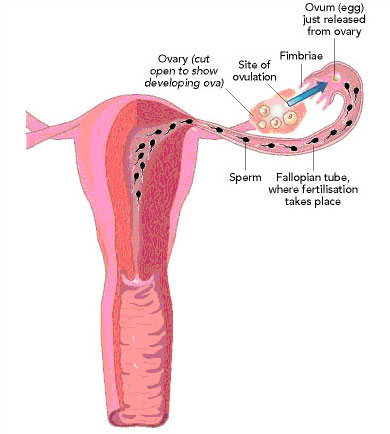
The fallopian tubes, or oviducts, consist of two tubes approximately 10 cm long that lead from the uterus and end in finger-like projections called fimbriae. The fimbriae ‘hover’ over the ovaries but are not attached to them.
During ovulation, the fimbriated end of the fallopian tube receives the mature ovum that is released from the ovary. The interior environment of the fallopian tube is biochemically complex. The ovum remains in the fallopian tube for a few days. Fertilisation normally takes place at the distal end of the fallopian tube, as can be seen in the figure. In tubal factor infertility problem, this tube is fully blocked resulting in permanent infertility.
If fertilisation occurs, the resulting embryo is held in the fallopian tube until it has developed into a small cell mass (blastocyst). It is then propelled through the fallopian tube by a combination of rhythmic contractions of the muscular walls of the tube (similar to the peristaltic muscular contractions of the gut), and the action of tiny hair-like projections called cilia. The embryo is swept toward the uterus where pregnancy may be established via implantation.
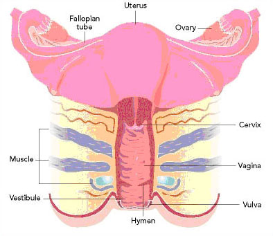
The uterus is a pear-shaped organ capable of undergoing major changes during a woman’s reproductive life. From puberty to the menopause, the inner lining of the uterus (the endometrium) provides a suitable environment for embryo implantation and development during pregnancy. The endometrial lining thickens during the proliferative phase (first half) of the menstrual cycle. It forms secretory glands after ovulation as it is stimulated by hormones released from the ovaries.
If the egg is not fertilized, or implantation does not occur, the endometrium is shed and excreted from the body via the vagina during menstruation and is slowly replaced in the course of the next menstrual cycle. The uterus also undergoes powerful, rhythmic contractions during labour, resulting in the delivery of the foetus at birth.
The uterus is composed of two main parts:
- Exclusively with the female in about 30 – 40% of cases.
- The narrow lower section called the neck, or cervix.
The upper portion of the uterine body is called the fundus. The fallopian tubes open into the opposite corners of the fundus and the cervix opens into the vagina. The cervix is a cylinder-like structure that leads from the upper end of the vagina into the interior of the uterus. It is about 2.5 cm long and has a fine canal running through it with openings at each end called the internal and external os, respectively. The inner walls of the cervix contain small sacs called crypts that secrete alkaline mucus, which protects sperm from the acidity of the vaginal secretions. The crypts also act as reservoirs for sperm.
The walls of the uterus contain three layers:
- The inner lining, called the endometrium.
- The middle muscular layer, called the myometrium.
- The outer serosal layer, called the peritoneum.
The serosa secretes a watery (serous) fluid that prevents friction between the uterus and surrounding organs. A cross-sectional view of the uterine structures is shown below.
The vagina is a tubular organ that extends from the cervix to its external opening. It is situated between the rectum, which lies beneath it, and the bladder and urethra, which lie above it.
It is composed primarily of smooth muscle lined with mucous membrane and functions to receive semen from the male. It also serves as the lower part of the birth canal and to act as the excretory duct for uterine secretions and menstrual flow.
The vulva is comprised of the external female genitals that surround the opening to the vagina and lubricate the passageway.
Vulvar structures include:
- The mons pubis, a skin-covered pad of fat over the pelvic joint that is covered by coarse hair in the adult female.
- The clitoris, an erectile structure similar to the penis in males.
- The labia minor, which is composed of fatty tissue and surrounds the opening of the vagina.
- The labia major, which contains hair follicles and sweat and sebaceous glands; the labia major is modified on the inner surface by the labia minor, which is moist and does not contain hair follicles.
- Bartholin’s glands, two bean-shaped glands located on either side of the vaginal opening that secrete lubricating fluid.
The breasts contain milk-producing glands and provide nourishment to the newborn. After birth, milk production is initiated and maintained by secretion of the hormone prolactin from the pituitary gland. The breasts are not fully developed in the female until well beyond the onset of menstruation. They are present in children and men only in rudimentary form.
Structures of the female reproductive system |
|||||||||||
| Structure | Description | Function | |||||||||
|---|---|---|---|---|---|---|---|---|---|---|---|
| Uterus | Pear-shaped cavity composed of the fundus, containing endometrial tissue, and a lower portion called the cervix. |
Site of embryo implantation and development. Provides muscular contractions to deliver foetus during labour. |
|||||||||
| Ovaries | Two almond-shaped structures located on the opposite sides of the pelvic cavity. Considered essential sex organs. |
Produce and store gametes (ova). Synthesise and release oestrogens and progesterone. |
|||||||||
| Fallopian Tubes | Ducts that end in finger-like projections (fimbriae) that hover over, but are not attached to, the ovaries. |
Ova pass through tubes from ovaries to uterus. Site of fertilisation. |
|||||||||
| Vagina | Canal leading from outside of body to cervix. |
Serves as lower part of birth canal. Receives sperm from male. |
|||||||||
| Vulva | Collective term for external genitalia (e.g. clitoris and labia). |
Surround and lubricate opening to vagina. |
|||||||||